Pretrain
38 datasets
Training and validation partitions supply the bulk of pretraining data.
SonoCorpus + SonoBase
1 Mohamed bin Zayed University of Artificial Intelligence, Abu Dhabi, UAE
2 Sheikh Tahnoon Bin Mohammed Medical City (STMC), Al Ain, UAE
3 King's College Hospital London - Dubai, Dubai, UAE
4 ADIA Lab, Abu Dhabi, UAE
* Correspondence: chao.qin@mbzuai.ac.ae, shadab@alumni.harvard.edu
Preprint: arXiv:2609.19230. Code MIT, weights CC BY-NC 4.0, data manifest CC BY 4.0.
The paper
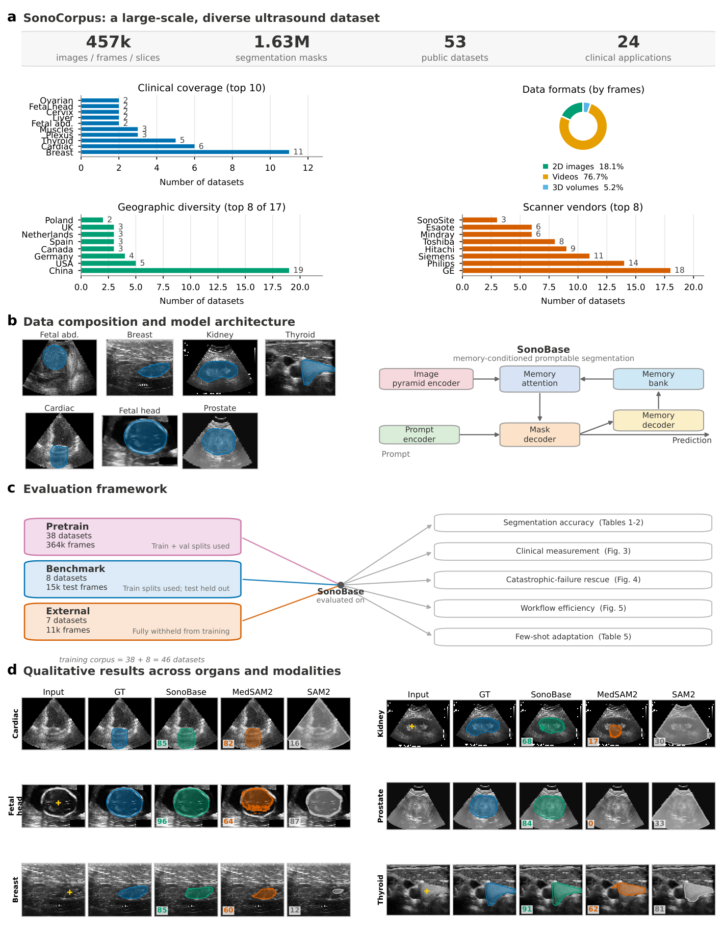
Ultrasound is the most widely deployed imaging modality worldwide, yet clinical AI remains fragmented into narrow single-task models that fail when device, operator, or anatomy changes. Here we present SonoCorpus, an open resource unifying 456,963 images and 1,626,085 expert masks from 53 public datasets spanning 24 clinical applications and 17 countries, and SonoBase, an interactive segmentation foundation model pretrained on it. Across fifteen evaluation datasets introducing new organs, devices, operators, and geographies, SonoBase outperforms other baselines (SAM2, SAM3, MedSAM2, etc.) on every dataset; on fully external data it exceeds the accuracy baselines achieve on their own in-distribution benchmarks. Ejection fraction derived from its segmentations falls within inter-observer variability (6.63% error), with fewer misclassifications at the defibrillator-candidacy threshold than any baseline (13% versus 18–42%); fetal head-circumference (1.81 mm) and gestational-age (1.2 days) errors fall below inter-observer variability. Where baselines fail outright, about one in five test cases, SonoBase recovers a useful segmentation in 81% of them, including on portable probes operated by novices in under-resourced settings (Sierra Leone and Tanzania). Five labeled examples adapt it to a new domain, and the identical training recipe transfers to different backbone, locating the advantage in ultrasound-specific pretraining rather than any single architecture. To ensure reproducibility and enable the community to build on SonoBase as a platform, we release all checkpoints, optimizer states, data-split indices, deduplication hashes, and starter code.
The data foundation
SonoCorpus consolidates 53 public ultrasound datasets into a unified resource for pretraining and evaluation. The collection spans 24 clinical applications across cardiac, fetal, breast, thyroid, kidney, prostate, nerve, muscle, and vascular imaging, with data from 17 countries acquired on scanners from all major manufacturers.
Three data formats: 2D images, video sequences, and 3D volumetric ultrasound.
Pretrain
Training and validation partitions supply the bulk of pretraining data.
Benchmark
Held-out test partitions evaluate cross-modality generalization. Training partitions are included in pretraining.
External
Completely withheld from pretraining, with separate test and few-shot adaptation pools.
Counts and modality labels reproduce the supplied inventory. Licence text reproduces the audit for the versions used. Source links lead to the supplied source or licence evidence; some point to an API record or terms page.
Modality follows the paper: 40 image, 8 video and 5 volumetric datasets (EchoCP, MUP, RegPro, SegThy, TDSC-ABUS keep slice structure).
The inventory loads when JavaScript is available.
Scroll horizontally to view all columns.
| Dataset | Tier | Modality | Frames | Masks | Licence | Source / evidence |
|---|
Redistribution also depends on access requirements and upstream terms. See Downloads and licences.
The segmentation model
SonoBase adapts SAM2 with an image-pyramid hybrid encoder. A Hiera-B+ transformer branch supplies global context at low resolution; ConvNeXt-S and ConvNeXt-T branches preserve texture and boundaries at higher resolutions. Cross-branch attention exchanges information at each of four hierarchical stages, and stage-wise fusion produces a unified multi-scale feature pyramid.
For single images, the memory pathway is disabled; for videos and 3D volumes, the memory mechanism enables prompt-once segmentation where a single user interaction on one frame propagates to the entire sequence.
Evidence across settings
The evaluation is retrospective; prospective validation, scoped per application and clinical context, is the natural next step and is directly enabled by the released platform.
The platform
SonoCorpus consolidates 53 public ultrasound datasets into a unified resource for pretraining and evaluation. We curated metadata including acquisition site, scanner manufacturer and model, patient demographics (when available), and image quality annotations, enabling evaluation under controlled distribution shifts rather than ad hoc data splits.
SonoBase adapts SAM2 by replacing its image encoder with an image-pyramid hybrid encoder while retaining the prompt encoder, mask decoder, and memory module that enable interactive segmentation and temporal propagation.
Held-out tests and external shifts
On eight held-out benchmark datasets, SonoBase achieves 74.4 mIoU with a single point prompt and 78.4 mIoU with a bounding-box prompt, compared to 28.1/52.4 for SAM2 and 37.3/54.0 for MedSAM2 (point/box mIoU; paired values follow this order throughout).
On seven completely external datasets, SonoBase maintains strong performance: 64.5 mIoU (point) and 79.8 mIoU (box), compared to 34.4/71.5 for MedSAM2 and 27.7/73.1 for SAM2. External performance exceeds what baselines achieve even on in-distribution benchmark data, indicating that SonoBase generalizes to unseen acquisition conditions rather than memorizing training-distribution shortcuts.
mIoU: mean intersection over union, reported on a percentage scale. Paired values above are point / box.
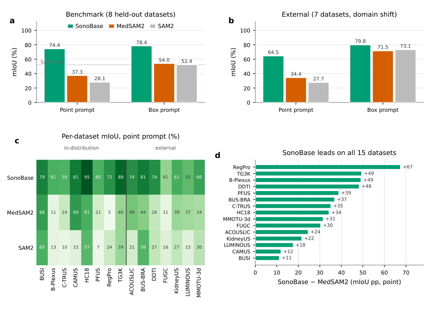
Figure image fig2_generalization.png could not be loaded.
A reusable training platform
To demonstrate it, we retrained the complete SonoBase recipe, the same 46 training datasets with the exact same data splits and the same schedule and loss, on SAM3.1 base, producing SB-SAM3, and evaluated all models under a matched box-prompt protocol scored by a single Dice implementation.
After identical fine-tuning on SonoCorpus both improve dramatically, and the SAM2-based SonoBase remains ahead: 0.897 versus 0.869, leading on 13 of 15 datasets.
Throughout, “SonoBase” refers to the released SAM2-based model; SB-SAM3 is a controlled comparison, not a second released model.
| Model | Ultrasound adaptation | Macro Dice |
|---|---|---|
| SAM2 | None | 0.823 |
| SAM3.1 | None | 0.823 |
| SB-SAM3 | Frozen encoder | 0.861 |
| SB-SAM3 | Full fine-tuning | 0.869 |
| SonoBase | Full fine-tuning | 0.897 |
Matched per-frame evaluation; memory propagation withheld. This protocol differs from the propagated segmentation benchmarks.
Because MedSAM3 cannot be driven by a box alone, it received its native text-plus-box prompt — strictly more information than the box-only SonoBase.
From contours to measurements
On the HC18 dataset (n = 201 images), SonoBase head circumference (HC) error measured 2.49 mm (point prompt) and 1.81 mm (box prompt), compared to a ground-truth ellipse-fitting floor of 1.37 mm.
SonoBase-derived GA estimates showed 1.20-day mean absolute error (box prompt) and 1.65 days (point), with 93.0% and 85.1% of cases within 3 days of true GA.
Ejection fraction: The box-prompt error falls within the reported inter-observer variability of 5.9–6.9% for echocardiographic EF measurement; the point-prompt error exceeds it.
Abdominal circumference on ACOUSLIC: With box prompts, the three models fall within 2.7 mm of one another and MedSAM2 records the lowest mean error, but the difference between SonoBase and MedSAM2 is not statistically significant (Wilcoxon p = 0.354), reflecting that a tight bounding box provides a strong geometric prior that narrows the gap between models.
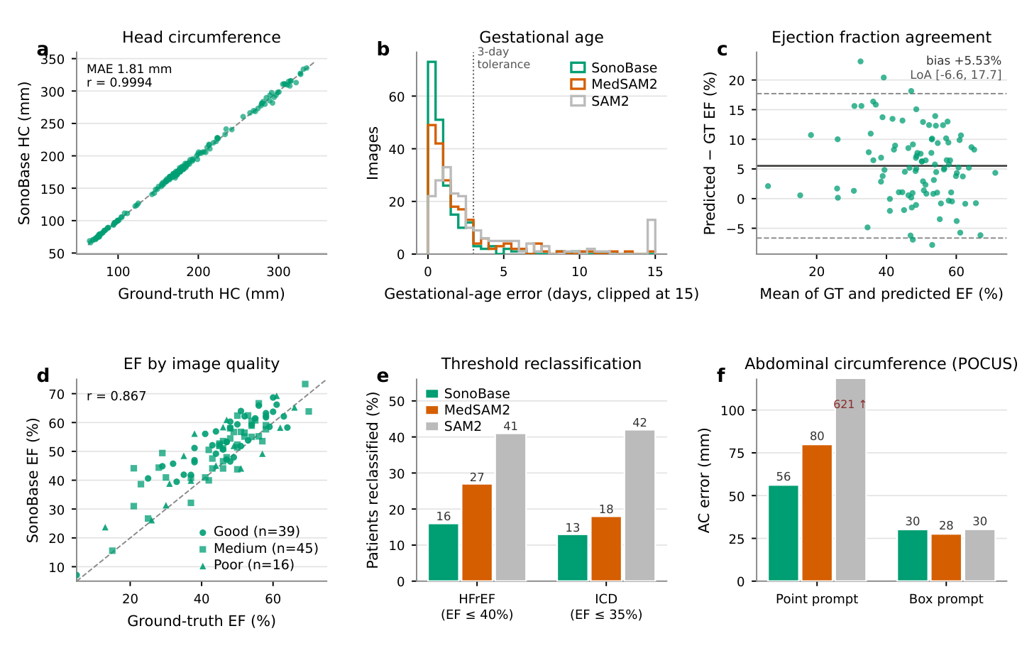
Figure image fig3_clinical.png could not be loaded.
Difficult cases and interaction
We define a catastrophic baseline failure as a test case on which a baseline model scores IoU below 10 under point prompting, effectively no usable segmentation; failure status is determined by the baseline scores alone. Across the 5,223 test cases of the fifteen evaluation datasets, at least one baseline failed catastrophically on 1,324 cases (25.3%). SonoBase recovered a clinically useful segmentation (IoU above 50) on 1,073 of those 1,324 cases (81.0%), corresponding to 20.5% of all test cases.
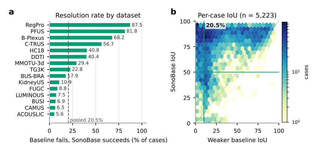
Figure image fig4_failure.png could not be loaded.
Side-by-side qualitative comparisons across datasets are Figure 4 of the paper; they are not reproduced here because each panel carries its own dataset licence and attribution requirements. The gallery below shows examples from datasets that permit redistribution with attribution.
On external datasets with box initialization, SonoBase starts at 79.8 mIoU and crosses the threshold with a single corrective click (6 seconds of clinician time); MedSAM2 needs five clicks (14 seconds) and SAM2 never crosses at any tested budget.
Usability threshold: 80% mIoU. Corrections follow an oracle protocol; clinician times are estimates from the Methods.
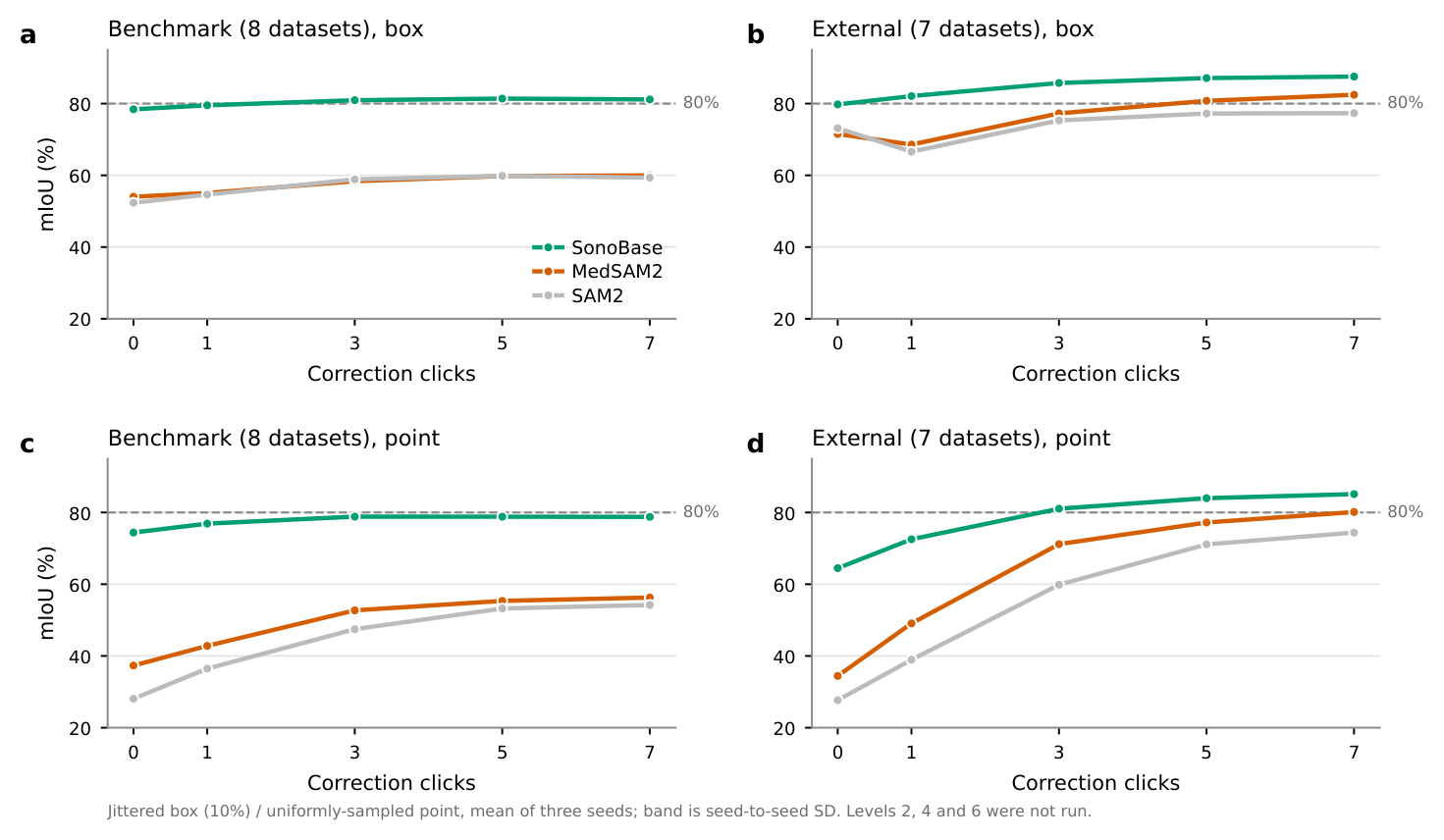
Figure image fig5_workflow.png could not be loaded.
Beyond the segmentation objective
Using SonoBase's encoder as a frozen backbone for US-RF-DETR (a detection and instance segmentation framework), SonoCorpus pretraining yielded 54.5 macro detection mAP across eight datasets, against 36.5 for SAM2 and 34.3 for MedSAM2, leading on all eight.
On a mouse brain tumor dataset (n = 1,203 frames), SonoBase achieved 47.0/64.2 mIoU (point/box), compared to 8.5/46.1 for SAM2 and 21.5/45.3 for MedSAM2.
Box-prompted performance is near-invariant to image quality (2.0 pp) and scanner manufacturer (1.4 pp); point-prompted performance is not (42.7 pp across manufacturers), so invariance claims are prompt-dependent.
mAP: mean average precision. pp: percentage points.
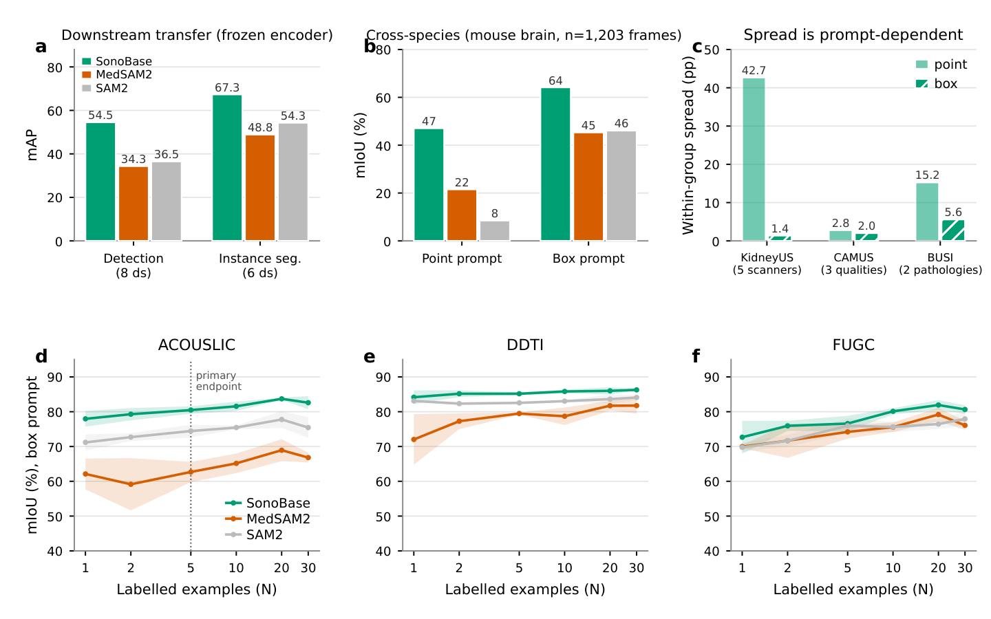
Figure image fig6_breadth.png could not be loaded.
Learning from a new domain
Adaptation updates the mask decoder; the image encoder, prompt encoder, and memory modules remain frozen.
The primary endpoint was met: at N = 5 with box prompts on ACOUSLIC, SonoBase reached 28.30 ± 6.92 mm AC MAE against 62.39 ± 42.55 mm for identically adapted MedSAM2 (p = 1.6 × 10⁻³⁰), converting the one zero-shot clinical endpoint on which a baseline led into a decisive advantage.
Adaptation saturates by N = 5–10 across models and datasets; practically, a site that labels five to ten cases captures nearly all of the available benefit.
Segmentation gains do not fully propagate to derived scalar measurements: against fine-tuned SAM2 the same AC endpoint is not significant (26.28 ± 3.05 mm, p = 0.051), because ellipse fitting absorbs much of the mask-quality difference.
Interactive segmentation
Upload an ultrasound image or pick an example, add positive and negative points or draw a box, and inspect the predicted mask. The Video tab propagates a first-frame prompt through a short clip.
The demo runs the released checkpoint on a Hugging Face Space. It is a research tool, not a medical device: upload only de-identified images you are authorised to process. If the embedded window does not load, open the Space directly.
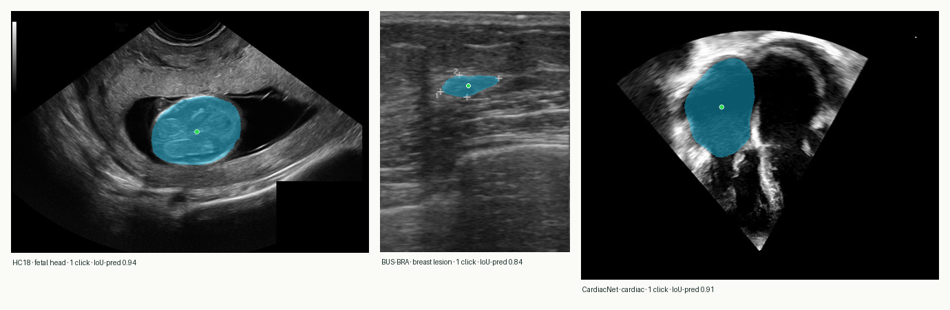
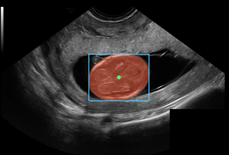
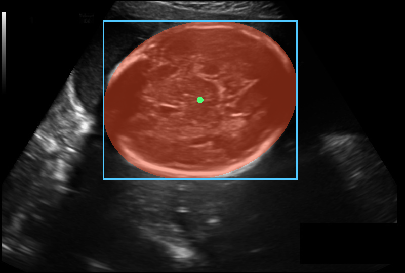
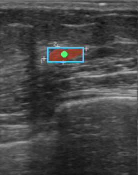
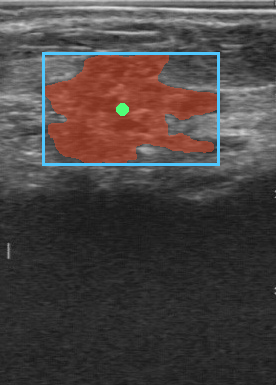
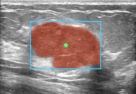
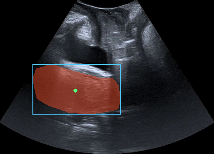
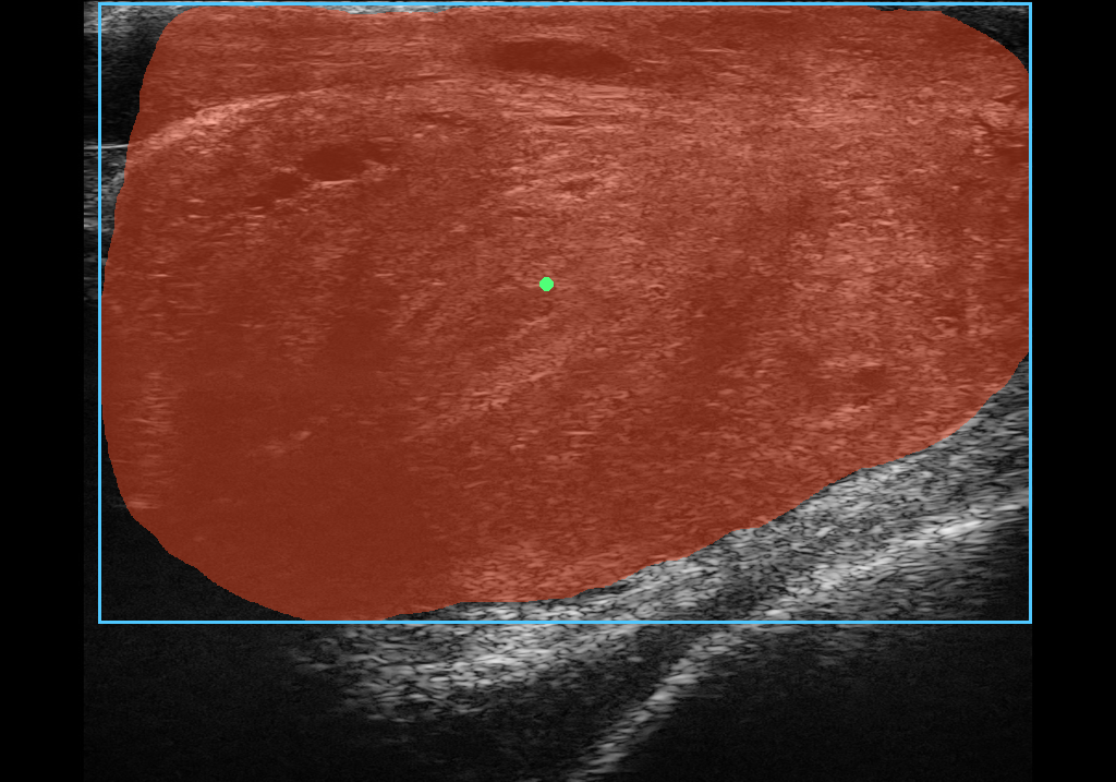
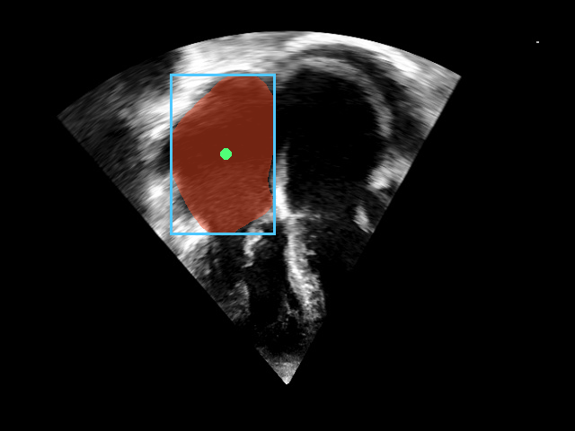
Click a tile to see the expert annotation (mask overlay, suggested point and box). All shown datasets permit redistribution with attribution; the full licence record is in the inventory.
Build on the platform
Checkpoints, optimizer states, stored data split indices, deduplication hashes, and starter code support reproduction and continued development.
SonoCorpus is not redistributed. The Zenodo record is a versioned manifest (CC BY 4.0): for each of the 53 datasets it gives the source page, the access conditions and licence as stated by the source, the exact split lists, and per-file checksums so that a copy obtained from the source can be verified against the one used in the study. Datasets are downloaded directly from their sources under their own terms; the manifest is kept up to date under a concept DOI, with each version frozen.
CAMUS and KidneyUS are link-only because access is registration-gated. BUS-UC is link-only because of conflicting upstream terms. BUSI is link-only because its dataset page does not state a reuse licence, even though the source article is openly licensed.
Code is released under the MIT License. Model weights and optimizer states are released under CC BY-NC 4.0 (non-commercial), because several constituent datasets of SonoCorpus are non-commercial.
Review the dataset licence inventory ↑Reference the work
Preprint citation; the author list matches the arXiv submission.
@article{SonoBase,
  title = {Open ultrasound foundation model for robust segmentation and clinical measurement across heterogeneous settings},
  author = {Qin, Chao and Khan, Fahad Shahbaz and Khan, Salman and Ather, Sarim and Anwar, Siddiq and Anwer, Rao Muhammad and Khan, Shadab},
  journal = {arXiv preprint arXiv:2609.19230},
  year = {2026},
  archivePrefix = {arXiv},
  eprint = {2609.19230},
  primaryClass = {cs.CV},
  doi = {10.48550/arXiv.2609.19230},
  url = {https://arxiv.org/abs/2609.19230}
}The work was funded by ADIA Lab and MBZUAI through internal research grants.